Understanding Your Lumbar MRI: Common Findings Explained
What Does a Lumbar MRI Show?
A lumbar MRI is designed to look closely at the structures of the lower back. In a standard study, the radiologist can evaluate:
- the vertebral bodies, meaning the blocks of bone that stack to form the spine
- the intervertebral discs between those bones
- the spinal canal, the main channel running down the middle
- the neural foramina, the openings where nerve roots exit
- the regions where the nerve roots travel
- the facet joints at the back of each level
- the ligaments and surrounding soft tissues
- the bone marrow and the vertebral endplates
That is a lot of detail, which is part of why reports feel overwhelming. Each of those structures gets described, level by level.
What Does a Lumbar MRI Not Show Well?
A standard lumbar MRI is set up to study the lumbar spine. Surrounding anatomy may appear at the edges of the images, but the study is not a dedicated examination of:
- the hips
- the entire pelvis
- the sacroiliac joints
- the abdominal or pelvic organs
This matters because low back, buttock and groin symptoms can come from more than one place. If your symptoms suggest a problem in one of those other areas, dedicated imaging or a different type of evaluation may sometimes be appropriate.
Does a lumbar MRI show the hips?
Parts of the hips may appear at the edge of some lumbar MRI images, but a lumbar MRI is not a dedicated hip examination.
Does a lumbar MRI show the pelvis?
Some pelvic structures may be partially visible, but a lumbar MRI is not designed as a complete pelvic MRI.
Does a lumbar MRI show the SI joints?
The sacroiliac joints may be partially visible depending on the study, but a standard lumbar MRI is not a dedicated SI-joint examination.
How to Read Your Lumbar MRI Report Without Getting Overwhelmed
You do not need to understand every sentence in the report. Five steps will get you most of the way there.
1. Start With the Impression
The impression is the radiologist's summary of what they consider the most important findings. If you read nothing else, read this. The long body of the report is the detailed support for that summary.
2. Find the Spinal Level
The lumbar levels are L1-L2, L2-L3, L3-L4, L4-L5 and L5-S1. Each part of the report usually describes what is happening at one of those levels, working down the spine. The level is an address, not a diagnosis.
3. Identify the Structure Being Described
Within each level, ask what the sentence is actually talking about:
- the disc
- the spinal canal
- the lateral recess
- the neural foramen
- the facet joint
- a nerve root
- a vertebral endplate
4. Look at the Relationship to the Nerve
Reports use words such as contact, abutment, crowding, displacement, impingement and compression to describe how close something sits to a nerve. These are not a fixed universal severity ladder, and different radiologists use them somewhat differently. They describe an anatomical relationship. The wording is compared in nerve-root abutment, impingement and compression.
5. Ask Whether It Matches Your Symptoms and Exam
This is the step that turns a report into useful information. Does the finding match:
- where your symptoms are
- how your pain travels
- your numbness or tingling
- any weakness
- your reflex findings
- what you can and cannot do
- the rest of your neurological examination
When those line up with the imaging, the finding becomes clinically convincing. When they do not, the finding may simply be part of the background anatomy.
The Basic Structure of a Lumbar MRI Report
Most lumbar MRI reports follow the same skeleton. There is a short section naming the study and technique, a comparison to any prior imaging, a few general observations about alignment and the spinal cord or cauda equina, and then the part patients usually fixate on: a level-by-level description.
The lumbar spine has five vertebrae, and the discs between them are named for the bones above and below. Going down the spine, the levels are L1-L2, L2-L3, L3-L4, L4-L5 and L5-S1. The radiologist walks through them one at a time and describes what is present at each.
L4-L5 and L5-S1 appear in reports constantly, which is why patients often assume those labels mean something is wrong. They do not. They are anatomical addresses. Those two levels carry the most load and move the most, so they are simply where degenerative change tends to show up first. A finding at L5-S1 matters when it lines up with where your symptoms actually are.
Why MRI Images Are Viewed in More Than One Direction
MRI produces slices through the body in different planes, and each plane answers a different question.
- Sagittal images are side views. They show the stack of vertebrae and discs from front to back, which is useful for judging disc signal, disc height, alignment and how far a disc extends backward.
- Axial images are cross sections, as if you were looking up through the spine from below. They show the shape of the spinal canal, the position of the nerve roots and whether something sits to the right or left of midline.
Some findings are much easier to appreciate in one plane than the other. Foraminal narrowing is often best judged on sagittal images, while the available space inside the central canal is usually clearer on axial images. This is one reason a single screenshot from a scan is a poor basis for conclusions. The radiologist reads the full series.
Common Disc Findings
Disc language is the most misread part of a report. These words describe water content, shape and position. They are descriptive, not diagnostic.
Disc Desiccation
Disc desiccation means the disc holds less water than a young healthy disc does, which makes it appear darker on T2-weighted images. It is a hallmark of ordinary disc degeneration and is extremely common with age. It does not mean you are dehydrated, and it does not mean the disc is the source of your pain. If your report mentions it, the useful next questions are which level, whether disc height has changed and whether anything is affecting a nerve. There is a full explanation in what disc desiccation means on an MRI.
Disc Bulge
A bulge describes a broad, generalized extension of the disc contour beyond the edges of the vertebral bodies, typically involving more than a quarter of the disc circumference. Because it is broad rather than focal, a bulge is not the same thing as a herniation, and calling every bulge a herniated disc creates unnecessary alarm. Bulges are common findings in people with no symptoms.
Disc Protrusion
A protrusion is a focal displacement of disc material. In standardized nomenclature, the base of the displaced material is wider than the part sticking out. It is one form of disc herniation in the technical sense of that word, but it is a localized shape change, and it can exist without symptoms.
Disc Extrusion
An extrusion is also a focal displacement, but the displaced material extends further than the width of its base. In some cases a fragment separates from the parent disc, which reports call a sequestration or a free fragment. Extrusion sounds more dramatic than protrusion, and it can be more likely to interact with nearby nerve structures, but it does not automatically mean surgery. Extrusions can and often do improve over time, and management still depends on symptoms, neurological findings and how the situation progresses.
Why the Size of a Disc Extrusion Is Only Part of the Story
Disc size is only one part of MRI interpretation.
A larger extrusion is not automatically more symptomatic than a smaller one, and two disc extrusions of similar size can affect surrounding structures differently depending on their location, direction of migration, available canal space and relationship to a nerve root.
Here is a useful example from a lumbar MRI containing disc extrusions at three different levels.
At L2-L3, the report described a broad-based disc extrusion with moderate canal stenosis, but no nerve-root impingement.
At L3-L4, the extrusion was considerably larger and severely migrating, with a possible sequestration component and a high probability of impingement of the descending right L4 nerve root. A sequestrated disc fragment refers to disc material that has lost continuity with the parent disc; in this study, the radiologist noted that a possible sequestration component at L3-L4 could not be excluded.
At L4-L5, another central extrusion indented the front of the thecal sac and crowded the descending L5 nerve roots.
The important point is that the measurement alone does not tell the whole story. The direction of the disc material, the amount of available space and its relationship to the nearby nerve roots all matter. Notice, too, how the report's language shifts with the anatomy: no nerve-root impingement detected at one level, crowding of nerve roots at another, and a high probability of impingement at a third. The radiologist also noted transitional anatomy at the lumbosacral junction, meaning level numbering required a defined convention for this examination.
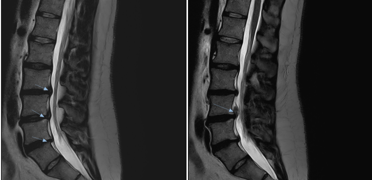
This is why MRI findings have to be matched to the patient's symptoms and neurological examination. A large extrusion may be important, but the clinical question is whether the affected level and nerve root match the patient's pain pattern, sensory findings, strength, reflexes and function.
Annular Fissure
The annulus fibrosus is the tough layered outer wall of the disc. An annular fissure is a separation between those fibers. Some reports still use the older phrase annular tear, but standardized nomenclature prefers fissure because tear implies an injury event that imaging cannot confirm. A fissure does not prove that anything happened to you. See the difference between an annular tear and an annular fissure for a longer discussion.
Disc Height Loss
As a disc degenerates it can lose height. That matters less as a standalone finding and more because of what it changes around it. When a disc flattens, the geometry of the nerve exit openings and the facet joints at that level changes too, and reports frequently mention height loss alongside foraminal narrowing or facet changes at the same level.
Can a Herniated Disc Change or Remodel Over Time?
Yes. A lumbar disc herniation is not necessarily a permanent, frozen finding.
This is well documented in the research literature rather than resting on individual stories. A systematic review of spontaneous regression of lumbar herniated discs and a later meta-analysis both found that a substantial proportion of herniations decrease in size over time without surgery. Extruded and sequestered disc herniations appear to have a greater tendency to regress than simple disc bulges.
Over time, herniated disc material can stay similar, enlarge, shrink or partially resorb.
That does not mean every herniation will improve without surgery, and it does not mean every patient should avoid surgical consultation. It does mean that the appearance of a disc on one MRI does not necessarily tell us what that disc will look like months later.
A Real-World Example of Disc Remodeling
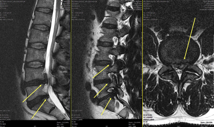
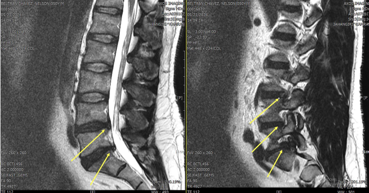
Here is one real-world example from our clinic.
The initial MRI described a very large L4-L5 disc extrusion extending approximately 22 mm along the back of the L4 vertebral body, with associated nerve-root impingement and canal narrowing. A measurement like that can sound alarming, but the size of an extrusion alone does not establish what care a patient needs.
On follow-up MRI, the L4-L5 extrusion was described at approximately 10 mm in craniocaudal extent, and the radiologist reported significant improvement/resorption of the central annular abnormality.
During the interval between these MRI studies, the patient underwent a course of conservative care that included non-surgical spinal decompression, low-level laser therapy and ARPwave.
Because this is a single clinical case, the imaging change cannot establish how much of the remodeling resulted from treatment versus the disc's natural biological resorption process. Other degenerative and foraminal findings remained on follow-up imaging; the educational point here is the documented remodeling of the large L4-L5 extrusion, not a claim that the entire MRI normalized.
Does a Large Disc Herniation Automatically Mean Surgery?
No. The size of a disc herniation on MRI alone does not determine whether surgery is required.
Treatment decisions should consider the whole clinical picture, including:
- symptom severity
- motor weakness
- progression of neurological deficits
- reflex changes
- sensory changes
- bowel or bladder dysfunction
- saddle-area numbness
- walking and standing tolerance
- functional limitations
- symptom duration
- response to appropriate conservative care
- the complete MRI findings
- patient goals
- medical or surgical consultation when appropriate
Surgery can be an appropriate option, and in some situations, such as progressive weakness or loss of bowel or bladder control, surgical evaluation can be urgent. A severe-looking MRI does not automatically mean that the spine has lost the ability to change. Some disc extrusions can remodel substantially over time. The more important clinical question is how the imaging fits with the patient's symptoms, neurological findings, function and response to care.
How This Fits With Published Research
The strongest evidence that lumbar disc herniations can shrink comes from systematic reviews and a meta-analysis of spontaneous regression, not from any single clinic's experience. Those reviews establish the biology: this can happen.
As additional clinical context, Dudum and Gatterman reported a four-patient case series in which patients received multimodal conservative care centered on non-surgical spinal decompression, with follow-up MRI showing measurable reductions in herniation size. A four-patient case series is low-level evidence. It does not show that spinal decompression causes disc resorption, and the protocol in that paper was not identical to the care used in the case above.
Spinal Narrowing and Nerve Findings
What "Stenosis" Means
Stenosis simply means narrowing of a space. The word tells you nothing until you know which space is narrowed, because the lumbar spine has several distinct ones, and they affect different nerves in different ways.
Central Canal Stenosis
The central canal is the main channel running down the middle of the spine. In the lumbar region it carries the cauda equina, the bundle of nerve roots below the end of the spinal cord. Central narrowing can come from disc material, thickened ligament, facet enlargement or a combination. When it is clinically significant, patients often describe leg heaviness, aching or fatigue that builds with walking or standing and eases when they sit or lean forward.
Lateral Recess Stenosis
The lateral recess is the side channel a nerve root travels through just before it reaches its exit opening. It is a separate region from both the central canal and the foramen, and narrowing here tends to affect a single traversing nerve root rather than the whole bundle.
Neural Foraminal Narrowing
The neural foramen is the opening on each side of the spine where a nerve root exits. Narrowing of that opening is described as foraminal stenosis or neural foraminal narrowing, and it is graded as mild, moderate or severe. Those grades are not as standardized between radiologists as patients assume, which is covered in the article on neural foraminal narrowing.
What Nerve Root Language Means
When a disc or bone spur sits near a nerve root, reports describe the relationship with words such as contact, abutment, impingement, displacement or compression. Patients naturally read these as a severity scale climbing from mild to severe.
They are not a formal severity scale. There is no universal, agreed definition separating each of those words, and different radiologists use them somewhat differently. They are descriptions of an anatomical relationship, and their meaning has to be interpreted alongside the images and your examination. The nuances are worked through in nerve root abutment, impingement and compression compared.
Why MRI Reports Often Contain Several Findings at Once
Lumbar MRI reports often contain more than one finding because the radiologist evaluates discs, vertebrae, joints, the spinal canal, neural foramina and nerve roots at every level.
In this real-world study, the report described annular fissures, several disc herniations, osteophytes, mild vertebral alignment changes, facet arthropathy, central stenosis, lateral recess narrowing, foraminal stenosis and nerve-root findings.
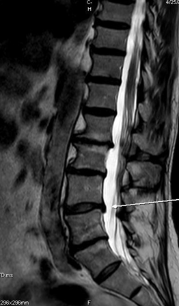
The presence of several MRI findings does not mean that every one of them is causing symptoms. Clinical interpretation requires determining which findings match the patient's history, symptom distribution, neurological examination and function.
Does a Large or Severe MRI Finding Automatically Mean Severe Symptoms?
No. Words such as mild, moderate and severe describe how pronounced an anatomical finding looks to the radiologist. They are not a rating of your pain.
"Severe" on an MRI describes the degree of an anatomical finding. It does not automatically mean the patient will have severe symptoms. Those grades do not by themselves describe disability, prognosis or the need for surgery.
The same is true of size. A large disc extrusion is not automatically more symptomatic than a smaller one. Where the disc material sits, which direction it has moved and how it relates to the surrounding nerve structures often matter more than the millimeter measurement.
Does an MRI Finding Automatically Explain Your Symptoms?
No. This is the single most important idea in this article.
Systematic reviews of imaging in people without back pain have found disc degeneration in roughly a third of adults in their twenties and in the large majority of adults by their sixties and seventies, along with high rates of disc bulges and other degenerative findings. Other work has found that degenerative findings are more prevalent in adults with low back pain than in asymptomatic controls. Both things are true at once: these findings are common in people who feel fine, and they are somewhat more common in people who hurt. That is exactly why presence alone cannot establish cause.
A finding becomes more clinically convincing when several things agree:
- the location of your symptoms
- the side your symptoms are on
- the neurological findings on examination, such as strength, sensation and reflexes
- what reproduces or relieves the symptoms during testing
- the level and side of the imaging finding
A simple example makes the point. If the MRI describes a right-sided protrusion at L4-L5 and your pain and numbness run down the left leg, that right-sided finding does not explain your left leg. Something else needs to account for it.
What Is Radiculopathy?
Radiculopathy is a clinical condition, not an imaging finding. It refers to dysfunction of a spinal nerve root, and it is identified from the pattern of symptoms and examination findings. Depending on the root involved, that can include radiating pain in a recognizable distribution, numbness or tingling, weakness in specific muscles, or a diminished reflex.
An MRI can support the diagnosis by showing something plausible at the matching level and side. It cannot establish radiculopathy on its own, and a report that uses the word compression is not the same as a clinical diagnosis of radiculopathy.
Bone, Joint and Degenerative Findings
- Degenerative disc disease. An umbrella phrase for age-related and wear-related disc change. Despite the word disease, it describes a process most people accumulate over time.
- Facet arthropathy. Degenerative change in the small paired joints at the back of each spinal level.
- Facet hypertrophy. Enlargement of those joints, often part of the same degenerative process, which can contribute to narrowing nearby spaces.
- Osteophyte. A bone spur, meaning extra bone formed along a joint or vertebral margin.
- Disc osteophyte complex. Disc material and bone spur formation described together because they blend into one contour on imaging.
- Spondylolisthesis. One vertebra sitting shifted relative to the one below it.
- Anterolisthesis. That shift going forward.
- Retrolisthesis. That shift going backward.
- Modic endplate change. Signal change in the bone directly above or below a disc, classified into types, often described alongside disc degeneration.
When Lower-Back or Leg Symptoms Need Prompt Medical Evaluation
Most degenerative findings are not urgent. A small number of symptom patterns are, and they deserve prompt medical evaluation regardless of what the imaging says:
- new loss of bowel or bladder control, or new difficulty urinating
- numbness in the saddle area, meaning the inner thighs, groin or buttocks
- weakness that is clearly getting worse over days rather than staying steady
- severe symptoms following significant trauma, such as a fall or a collision
- severe back pain with fever, unexplained weight loss or a history that raises concern for infection or cancer
- any finding your physician flags as needing medical or surgical assessment
To be clear about the opposite direction as well: a large herniation on a report is not automatically an emergency. Size on imaging and clinical urgency are different things.
What to Bring to a Spine Evaluation
The evaluation goes much further when the clinician can compare the report against a clear account of your symptoms. Useful things to bring:
- the radiology report itself, not just a summary you were told verbally
- the MRI images, on disc or through a patient portal, if you can get them
- any previous imaging, including older X-rays or MRIs, for comparison
- a written list of your symptoms in plain language
- which side and exactly how far down the leg symptoms travel
- when symptoms started and whether anything set them off
- positions and activities that make them worse or better
- what treatment you have already tried and how you responded
- a current medication list
- any surgical opinions you have already received
Explore Specific MRI Findings
Each of these articles goes deeper into one term you may see in your report:
- Disc desiccation on MRI, and why a darker disc on T2 images is so common with age.
- Annular fissure or annular tear on MRI, and why the wording changed.
- Nerve-root abutment, impingement and compression, and what those words do and do not mean.
- Neural foraminal narrowing, and how mild, moderate and severe grades are applied.
The Main Takeaway
The goal of reading a lumbar MRI is not simply to find the most alarming word in the report. The goal is to understand what structures are affected, where they are affected, and whether those findings match your symptoms and examination.
Your MRI report is a careful description of anatomy at a moment in time. It becomes useful when someone checks whether what the scan describes actually corresponds to what your body is doing.
Have a lumbar MRI and want help understanding how the findings relate to your symptoms? A clinical evaluation can help put the imaging into context and determine whether conservative care, additional medical evaluation or surgical consultation may be appropriate.
Frequently asked questions
References
- Fardon DF, Williams AL, Dohring EJ, et al. Lumbar disc nomenclature: version 2.0. Recommendations of the combined task forces of the North American Spine Society, the American Society of Spine Radiology and the American Society of Neuroradiology. Spine J. 2014;14(11):2525-2545. PMID: 24768732.
- Brinjikji W, Luetmer PH, Comstock B, et al. Systematic literature review of imaging features of spinal degeneration in asymptomatic populations. AJNR Am J Neuroradiol. 2015;36(4):811-816. PMID: 25430861.
- Brinjikji W, Diehn FE, Jarvik JG, et al. MRI findings of disc degeneration are more prevalent in adults with low back pain than in asymptomatic controls: a systematic review and meta-analysis. AJNR Am J Neuroradiol. 2015;36(12):2394-2399. PMID: 26359154.
- Chiu CC, Chuang TY, Chang KH, et al. The probability of spontaneous regression of lumbar herniated disc: a systematic review. Clin Rehabil. 2015;29(2):184-195. PMID: 25009200.
- Zhong M, Liu JT, Jiang H, et al. Incidence of spontaneous resorption of lumbar disc herniation: a meta-analysis. Pain Physician. 2017;20(1):E45-E52. PMID: 28072796.
- Dudum J, Gatterman B. Reduction of the size of a lumbar disc herniation using non-surgical spinal decompression combined with chiropractic care. J Contemp Chiropr. 2024;7(1):146-155. Four-patient case series.