Disc Desiccation on MRI: What It Means and When It Matters
What Is Disc Desiccation?
A healthy intervertebral disc holds a great deal of water, particularly in the nucleus at its center. That water content is maintained by large molecules called proteoglycans, which attract and hold fluid and give the disc its ability to distribute load.
With aging and degeneration, the composition of the disc changes. Proteoglycan content falls, the collagen structure changes, and the disc holds less water than it once did. On T2-weighted MRI images, where fluid appears bright, a disc with lower water content appears darker than its neighbors. That darker appearance is what gets reported as disc desiccation.
It is worth resisting the mental image of a disc drying out like a sponge left on a counter. What the imaging reflects is a slow change in the biology and structure of the tissue, not simple evaporation.
A Real-World Example of Multilevel Disc Desiccation
On T2-weighted MRI, a healthier lumbar disc generally contains more water and therefore tends to appear brighter. As a disc loses hydration, its signal may become darker.
In this MRI, several lumbar discs demonstrate reduced T2 signal, particularly in the lower lumbar spine, consistent with multilevel disc desiccation and degenerative disc change.
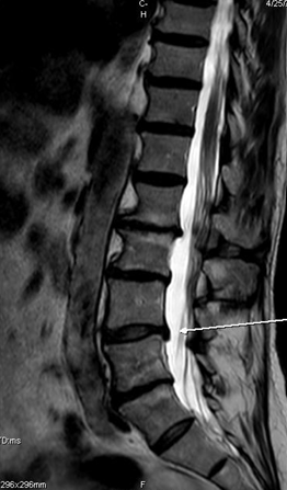
The accompanying radiology report focuses primarily on other findings, including disc herniations, annular fissures, osteophytes and spinal narrowing. This is a useful example of why the full MRI appearance can contain degenerative features that may not each be listed as a separate line item in the report.
Disc Desiccation vs Disc Height Loss
Disc desiccation and disc height loss are related but different findings. Desiccation refers to reduced water content within the disc. Disc height loss means the physical space between adjacent vertebral bodies has decreased. A disc can lose hydration before substantial height loss develops.
Disc Desiccation vs Disc Herniation
A desiccated disc is also not the same thing as a herniated disc. Desiccation describes the hydration and signal characteristics of the disc, while herniation describes displacement of disc material beyond its normal boundary. The two findings may occur together.
Disc Desiccation Rarely Appears in Isolation
This study also demonstrates that disc desiccation rarely needs to be interpreted in isolation. The same lumbar MRI may contain annular fissures, osteophytes, bulges or herniations, spinal and foraminal narrowing, alignment changes and nerve-root findings.
The presence of several degenerative findings does not mean that every one of them is producing symptoms. The plain-English guide to your lumbar MRI report explains how these separate descriptions fit together in one study.
Does Disc Desiccation Mean I Am Dehydrated?
No. This is probably the most common misunderstanding of the term, and it is an understandable one, since desiccation literally means drying.
Staying reasonably hydrated is good for you for many reasons. But the radiologist is not telling you to drink more water. Disc water content is governed by the tissue's own chemistry and by loading through the day, not by how many glasses you had this morning. There is no evidence that increasing fluid intake reverses disc desiccation or changes how a disc looks on a repeat MRI.
Is Disc Desiccation the Same as Degenerative Disc Disease?
They overlap, but they are not interchangeable. Degenerative disc disease is a broader label for the collection of changes a disc and its neighboring structures accumulate over time. Desiccation is one feature within that picture.
Disc degeneration can involve any combination of:
- reduced water content and darker T2 signal
- loss of disc height
- annular fissuring
- bulging or focal displacement of disc material
- endplate changes in the adjacent bone
- osteophyte formation
A report might mention desiccation alone at one level and several of these together at another. That difference is more informative than the word desiccation by itself.
Does Disc Desiccation Always Cause Pain?
No, and the research on this is fairly clear.
Systematic reviews of imaging in people with no back pain have found disc degeneration in roughly a third of adults in their twenties, rising to the large majority of adults by their sixties and seventies. Plenty of people walk around with desiccated discs and feel completely fine.
At the same time, other pooled analyses have found that degenerative disc findings are more prevalent in adults with low back pain than in asymptomatic controls, particularly in younger adults. So the finding is not meaningless either.
Holding both facts together leads to the correct conclusion: the presence of desiccation raises a possibility, it does not prove causation, and it never proves causation in one specific patient without clinical correlation.
When Does Disc Desiccation Matter More?
The word "desiccation" describes an imaging characteristic. It does not tell us by itself how much pain a patient should have or whether treatment is required.
Desiccation becomes more clinically interesting when the surrounding details start agreeing with the patient in front of you. Things a clinician weighs:
- where the symptoms actually are, and whether they stay local or radiate
- whether there is a mechanical pattern, meaning specific positions, loads or movements that reliably reproduce or relieve the symptoms
- whether there are neurological findings such as sensory change, weakness or reflex change
- whether the same level also shows a protrusion or extrusion
- whether disc height has been lost, and what that has done to nearby spaces
- whether central canal, lateral recess or foraminal narrowing is described
- whether a nerve root is described as involved, and on which side
- what the orthopedic and neurological examination shows
A single desiccated disc at L4-L5 in a patient whose examination is unremarkable means something very different from a desiccated disc with height loss and matching foraminal narrowing in a patient with a clear dermatomal symptom pattern on the same side.
Can Disc Desiccation Be Reversed?
This deserves a careful answer, because it is a place where patients are frequently promised more than anyone can deliver.
There is no treatment that has been shown to reliably restore normal water content to a degenerated disc or to return an MRI to a younger appearance. Claims that a particular table, laser, supplement or program rehydrates or regenerates discs go beyond what the evidence supports.
What responsible care does target is the clinical problem: symptoms, function, loading tolerance, and any mechanical or neurological findings that are genuinely relevant. People often improve a great deal without the imaging changing at all, which is a useful reminder that the MRI was never measuring how you feel.
Disc Desiccation vs Disc Bulge
These describe different properties. Desiccation is about signal, meaning water content. A bulge is about contour, meaning the disc extends beyond its normal margins in a broad, generalized way.
A disc can show:
- desiccation with a completely normal contour
- a bulge without producing any symptoms
- both together, along with height loss, at a level that is degenerating
Disc Desiccation vs Herniated Disc
Herniation is a focal event in shape. It refers to localized displacement of disc material beyond the normal boundaries of the disc space, and standardized nomenclature subdivides it into protrusion and extrusion based on the geometry of the displaced material.
Desiccation is not a herniation and does not become one. A degenerating disc may be more prone to changes in contour over time, but the two terms are describing different things, and the presence of one does not imply the other.
If your report describes both, the herniation description is usually the more clinically relevant half, and even then only if it matches your symptoms and examination. The comparison is laid out further in the article on annular fissures and how they differ from herniations.
What Should You Do If Your Report Says "Disc Desiccation"?
Bring the report to an evaluation and work through the questions that actually change the picture:
- Which level or levels are involved?
- Is there disc height loss at that level?
- Is a protrusion or extrusion described?
- Is any narrowing described, and of which space?
- Is a nerve root described as affected, and on which side?
- Does the side and level match where my symptoms are?
- Does my examination support that this level is involved?
For the wider vocabulary that shows up alongside desiccation, the guide to common lumbar MRI findings walks through the full report section by section.
Frequently asked questions
References
- Brinjikji W, Luetmer PH, Comstock B, et al. Systematic literature review of imaging features of spinal degeneration in asymptomatic populations. AJNR Am J Neuroradiol. 2015;36(4):811-816. PMID: 25430861.
- Brinjikji W, Diehn FE, Jarvik JG, et al. MRI findings of disc degeneration are more prevalent in adults with low back pain than in asymptomatic controls: a systematic review and meta-analysis. AJNR Am J Neuroradiol. 2015;36(12):2394-2399. PMID: 26359154.
- Fardon DF, Williams AL, Dohring EJ, et al. Lumbar disc nomenclature: version 2.0. Spine J. 2014;14(11):2525-2545. PMID: 24768732.