Annular Tear or Annular Fissure on MRI: What Does It Mean?
What Is the Annulus Fibrosus?
Each intervertebral disc has two main parts. The nucleus pulposus sits in the center and is the softer, more fluid-rich portion. Surrounding it is the annulus fibrosus, a wall built from concentric layers of collagen fibers, with each layer running at an angle to the next.
That crosshatched construction is what lets the disc handle compression, bending and twisting at the same time. The layers are anchored into the vertebral bodies above and below, and the outer portion of the annulus carries a nerve supply, which is part of why it is discussed in conversations about disc-related pain.
You will sometimes hear the disc compared to a jelly donut. The analogy captures the soft center and firmer outer ring, but it badly understates the annulus, which is a layered structural wall rather than a thin shell waiting to burst.
What Is an Annular Fissure?
An annular fissure is a separation between annular fibers, or between fibers and their attachment to the vertebral body. It can involve one layer or extend through several.
Radiology and anatomy literature classifies fissures by their orientation, describing concentric, radial and transverse patterns depending on how the separation runs through the annular layers. That taxonomy is useful for researchers and for radiologists comparing studies. For a patient reading a report, the more relevant point is simply that a separation is present in the outer part of the disc.
Why Do Some Reports Say "Annular Tear"?
Language in spine imaging has shifted over the last few decades. Annular tear was common in older reports and is still used by some radiologists out of habit or because it is the phrase referring clinicians recognize.
The standardized lumbar disc nomenclature developed jointly by the North American Spine Society, the American Society of Spine Radiology and the American Society of Neuroradiology recommends fissure specifically because tear carries an implication of injury. An imaging study cannot tell you that something tore. It can only tell you that a separation is visible now.
If your report says tear, it does not mean your radiologist believes you were hurt. It usually means the older word was used.
Does an Annular Fissure Mean the Disc Was Injured?
Not necessarily. Annular fissures are frequently associated with degenerative change in the disc, and degenerative change develops gradually and quietly in most people.
Trauma can certainly play a role in some cases, and the history matters. But the finding itself does not establish a mechanism or a date. Two patients can have the same description on their reports, one after a specific incident and one with no memorable event at all.
Is an Annular Fissure the Same as a Herniated Disc?
No. These describe different things.
- An annular fissure is a change within the fibers of the disc wall.
- A disc herniation is localized displacement of disc material beyond the normal boundaries of the disc space, which nomenclature further divides into protrusion and extrusion based on the shape of the displaced material.
They can coexist. Displaced material generally has to move through a defect in the annulus, so a report may describe both at the same level. But having a fissure does not mean you have a herniation, and the words are not interchangeable.
What Is a High-Intensity Zone?
A high-intensity zone is a small bright area sometimes seen within the posterior annulus on T2-weighted images, standing out against the darker surrounding annular tissue. It often corresponds with an annular fissure.
It is worth being careful here, because the high-intensity zone has been oversold at times as a marker of the painful disc. The relationship is not that clean. High-intensity zones are also seen in people without symptoms, and the reported association with discogenic pain varies considerably across studies. Treat it as one piece of information among many rather than a diagnosis in itself.
A Real-World MRI Example of Annular Fissures
On T2-weighted MRI, an annular fissure may sometimes appear as a small region of increased signal within the outer portion of a disc. Radiologists may describe this as a high-intensity zone.
In this study, the radiologist described zones of hyperintensity consistent with annular fissures at L2-L3, L3-L4 and L5-S1.
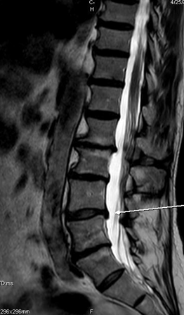
The same MRI also contained several other degenerative findings, including osteophytes, disc herniations, vertebral alignment changes, central and foraminal stenosis, facet arthropathy and nerve-root findings. This is important because an annular fissure should not automatically be interpreted in isolation.
Annular Fissures Often Appear in a Broader Degenerative Picture
This case also demonstrates that annular fissures may appear alongside broader degenerative findings. The same MRI report described osteophytes, retrolisthesis, facet arthropathy, disc herniations and several types of spinal narrowing, including neural foraminal narrowing at more than one level.
That does not mean degeneration automatically causes symptoms or progressive disability. It means that an MRI often contains several structural findings that must be considered together, which is exactly why the full lumbar MRI report walkthrough reads the study as a whole rather than one phrase at a time.
Does an Annular Fissure Always Cause Pain?
No. Annular fissures show up in imaging of people who have no back pain, and their prevalence increases with age alongside other degenerative findings.
There is research exploring associations between annular fissures and discogenic pain, and the innervation of the outer annulus offers a plausible mechanism. That is genuinely different from proving that your fissure is producing your symptoms. Imaging can identify a candidate. Only clinical correlation can make a candidate convincing.
Can an Annular Fissure Heal?
This is a fair question with an honest and slightly unsatisfying answer. Disc tissue has a limited blood supply and does not repair the way muscle or skin does, so it would be overpromising to say a fissure closes and disappears.
What does change is the clinical situation. Symptoms often settle over weeks to months, tissue behavior and irritability change, and people frequently return to activities they had given up. Care is aimed at that clinical trajectory rather than at chasing an MRI label, and repeating imaging simply to see whether a fissure looks different rarely changes what should be done.
What Makes an Annular Fissure More Clinically Relevant?
A fissure earns more attention when the rest of the picture agrees with it:
- a symptom pattern that is consistent and reproducible rather than vague and shifting
- other disc changes at the same level, such as desiccation or height loss
- an associated protrusion or extrusion
- described involvement of a nerve root on the side that matches the symptoms
- examination findings that point to the same level
- the absence of a more convincing competing explanation, such as hip pathology, sacroiliac involvement or a non-spinal source
That last point gets skipped often. A finding is only compelling when the alternatives have been considered.
Questions to Ask When Your MRI Says Annular Fissure
- Which level is it at, and which part of the annulus?
- Is there also a protrusion or extrusion at that level?
- Is any nerve root described as involved?
- Is there desiccation or disc height loss at the same level?
- Does the described location correspond with where my symptoms are?
- What did the examination find, and does it agree with the imaging?
- What would change management, and what would not?
For the rest of the vocabulary in your report, the plain-English walkthrough of a lumbar MRI report covers each section in turn, and disc desiccation is the finding most often listed alongside a fissure at the same level.
Frequently asked questions
References
- Fardon DF, Williams AL, Dohring EJ, et al. Lumbar disc nomenclature: version 2.0. Recommendations of the combined task forces of the North American Spine Society, the American Society of Spine Radiology and the American Society of Neuroradiology. Spine J. 2014;14(11):2525-2545. PMID: 24768732.
- Brinjikji W, Luetmer PH, Comstock B, et al. Systematic literature review of imaging features of spinal degeneration in asymptomatic populations. AJNR Am J Neuroradiol. 2015;36(4):811-816. PMID: 25430861.
- Brinjikji W, Diehn FE, Jarvik JG, et al. MRI findings of disc degeneration are more prevalent in adults with low back pain than in asymptomatic controls: a systematic review and meta-analysis. AJNR Am J Neuroradiol. 2015;36(12):2394-2399. PMID: 26359154.