Neural Foraminal Narrowing on MRI: What It Means
What Is the Neural Foramen?
Between every two adjacent vertebrae there is an opening on each side, called the neural foramen or intervertebral foramen. A nerve root passes through it as it leaves the spinal canal on its way to the leg.
The boundaries of that opening are formed by the disc and vertebral bodies in front, the facet joint behind, and the bony pedicles above and below. Because the disc forms part of the front wall, changes in disc height directly change the height of the opening.
What Does Neural Foraminal Narrowing Mean on MRI?
It means the radiologist judged the opening to be smaller than expected at that level and side. Reports typically identify the level, such as L4-L5 or L5-S1, note whether it is left, right or bilateral, and apply a descriptive grade such as mild, moderate or severe.
Some reports go further and comment on whether the exiting nerve root appears contacted, displaced or compressed within the narrowed opening. Those additional sentences usually carry more clinical information than the grading word does.
Foraminal vs Central Canal vs Lateral Recess Stenosis
Reports often describe more than one type of narrowing, and they are not the same thing. The distinction is about location:
- Central canal stenosis involves the main canal in the middle of the spine, where the nerve roots travel as a bundle below the end of the spinal cord.
- Lateral recess stenosis involves the side channel within the canal, where a nerve root travels shortly before turning to exit.
- Foraminal stenosis involves the exit opening itself, affecting the root leaving at that level.
Because different roots occupy these spaces at a given level, the location of the narrowing influences which root would be implicated if the finding turns out to be clinically relevant.
One Disc Level Can Affect Different Nerve Roots in Different Places
One reason lumbar MRI terminology can be confusing is that the same disc level can affect different nerve structures depending on where the narrowing occurs.
At L5-S1, for example, the exiting L5 nerve root travels through the neural foramen, while the descending S1 nerve root passes lower through the spinal canal before exiting at the next level. That means an L5-S1 disc problem can potentially affect the exiting L5 root, the descending S1 root, or both, depending on the location and direction of the disc material.
This MRI provides a useful real-world example.
The radiology report described a broad-based L5-S1 disc extrusion measuring approximately 12 x 19 x 7 mm. The report noted a high probability of impingement of the descending left S1 nerve root within the canal region, while also describing mild-to-moderate foraminal compromise with crowding and probable irritation of the exiting L5 nerve roots.
This is why terms such as central canal stenosis, foraminal narrowing and nerve-root impingement should not be treated as interchangeable. They describe different anatomical relationships. Central canal stenosis is narrowing of the main spinal canal; neural foraminal narrowing is narrowing of the opening where the exiting nerve root leaves the spine. Neither is inherently more serious than the other. Clinical significance depends on the severity of the narrowing, which nerve is affected, the neurological examination, the symptoms, the person's function and the overall anatomy.
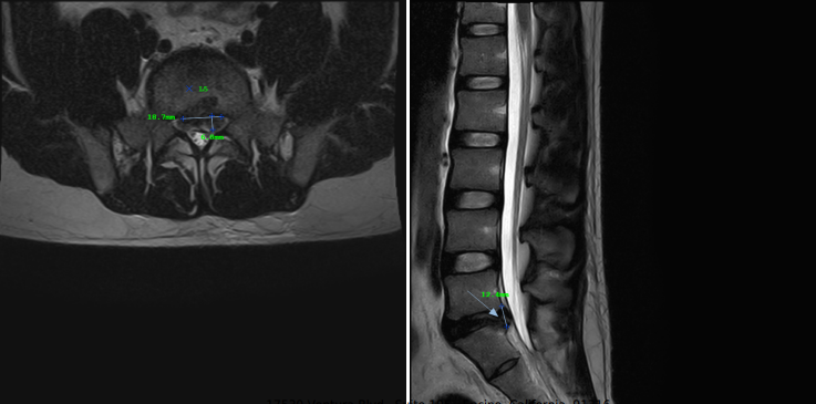
Exiting vs descending nerve roots
At L5-S1, the L5 nerve root exits through the neural foramen, while the S1 nerve root is still descending through the canal before exiting lower down. A disc problem at L5-S1 can therefore affect one or both nerve roots depending on where the disc material extends.
The important question is not simply whether an MRI says "stenosis." The more useful questions are where the narrowing is located, which nerve root is nearby, and whether the patient's symptoms and neurological examination match that anatomy.
What Causes Neural Foraminal Narrowing?
Common contributors include:
- loss of disc height, which lowers the roof-to-floor dimension of the opening
- disc bulging or a herniation extending into or near the foramen
- facet joint enlargement from arthritic change at the back of the opening
- thickening of the ligamentum flavum
- osteophytes, meaning bone spurs, forming along the joint or vertebral margins
- spondylolisthesis, where one vertebra shifts forward relative to the one below
Several of these frequently occur together, which is why a report may list foraminal narrowing alongside disc desiccation and height loss at the same level.
Foraminal Narrowing Can Occur at Multiple Levels
Some patients have foraminal narrowing at more than one lumbar level. In this MRI, the radiologist described neural foraminal narrowing from L2-L3 through L5-S1, ranging from mild to severe depending on the level and side. The same study also showed osteophytes, retrolisthesis, facet arthropathy and disc herniations.
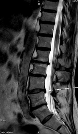
This illustrates how foraminal narrowing may develop as part of a broader degenerative pattern rather than from a single isolated structure. That same report also described annular fissures at several levels, and the plain-English lumbar MRI walkthrough explains how these separate descriptions fit together in one report.
Does Foraminal Narrowing Always Cause Symptoms?
No. Degenerative narrowing is common in imaging of people who have no back or leg symptoms, and its prevalence rises steadily with age.
The reverse is also true. Someone can have meaningful leg symptoms with only modest narrowing described, because symptoms depend on far more than a measured space, including inflammation, nerve sensitivity, position and load through the day.
How Positions and Movement Affect the Foramen
One thing a static MRI cannot show is that the foramen changes size as you move. Extending backward generally reduces the opening, while flexing forward generally opens it somewhat.
This is why many people with foraminal or central narrowing notice that symptoms increase with prolonged standing or walking upright and ease when they sit, lean forward or rest a hand on a cart. That pattern is clinically interesting because it is behavioral evidence pointing at the same mechanism the imaging described.
Your MRI was taken lying still in one position. Your symptoms happen across a whole day of positions. Both pieces of information matter.
What Do Mild, Moderate and Severe Mean?
These are descriptive grades that convey the radiologist's impression of how reduced the space appears. Grading systems for foraminal narrowing have been proposed and are used in research and in some practices, but no single standard is applied identically everywhere. Two radiologists may word the same anatomy slightly differently.
Two implications follow. First, the grade is not a treatment plan, and severe on a report does not automatically mean surgery. Second, a mild grade does not dismiss your symptoms. Clinicians weigh grades as one input among symptoms, examination findings and functional limitations.
When Is Foraminal Narrowing Clinically Relevant?
The finding gains weight when the rest of the picture agrees:
- the side of the narrowing matches the side of the symptoms
- the level plausibly corresponds to the symptom distribution
- examination findings point to the same nerve root
- symptoms behave in a way consistent with the mechanism, such as worsening with extension
- the report describes the exiting root as contacted, displaced or compressed
- competing explanations have been considered and do not fit as well
The language used about the nerve root in that same sentence deserves attention, and it is easy to over-read. What contact, abutment, impingement and compression do and do not mean is covered in the guide to nerve root terminology.
When to Seek Prompt Medical Evaluation
Most foraminal narrowing is not urgent. Seek prompt medical evaluation for:
- progressive weakness in a leg or foot
- new loss of bowel or bladder control
- numbness in the saddle area, meaning the inner thighs, groin or buttocks
- severe neurological symptoms in both legs
- severe symptoms following significant trauma
Making Sense of Your Report
Read the narrowing description together with the rest of the report rather than in isolation, then bring the questions that matter to an evaluation: which level, which side, which root, what the examination shows, and whether all of it points to the same place. The full walkthrough of a lumbar MRI report covers the remaining terminology, and annular fissures are another finding commonly listed at the same level.
Frequently asked questions
References
- Brinjikji W, Luetmer PH, Comstock B, et al. Systematic literature review of imaging features of spinal degeneration in asymptomatic populations. AJNR Am J Neuroradiol. 2015;36(4):811-816. PMID: 25430861.
- Fardon DF, Williams AL, Dohring EJ, et al. Lumbar disc nomenclature: version 2.0. Spine J. 2014;14(11):2525-2545. PMID: 24768732.