Nerve Root Abutment vs Impingement vs Compression on MRI
What Is a Spinal Nerve Root?
Nerves that supply your legs begin as roots inside the spinal canal. In the lumbar spine, those roots travel down through the canal, pass through a side channel called the lateral recess, and then exit through an opening on each side called the neural foramen. Once outside, they join with others to form the larger peripheral nerves, including the sciatic nerve.
Because each root serves a fairly predictable set of muscles and a general area of skin, a problem at a specific root tends to produce a recognizable pattern. That predictability is what lets an examination point toward a level.
What Does "Contact" Mean?
Contact means the structure touches the nerve root on the images, with no space visible between them at that point. It is the mildest of the descriptions in the sense of what it claims, because it claims very little.
Contact is not injury. Structures in the spinal canal sit close together, and touching happens routinely in people with no symptoms at all.
What Does "Abutment" Mean?
Abutment means the structure is immediately against the nerve root, pressed up alongside it. It is close in meaning to contact, and many radiologists use the two nearly interchangeably.
It would be a mistake to translate abutment as always mild. Some radiologists reserve it for situations where a structure is firmly against the root without obvious deformity, and others use it more loosely. The word narrows the possibilities. It does not settle them.
What Does "Impingement" Mean?
Impingement generally suggests that the structure is encroaching on the nerve root rather than merely resting against it, implying a more meaningful interaction. In practice, use varies widely. Some reports use impingement where others would say compression, and some use it where others would say abutment.
When this word appears, the useful move is to read the rest of the sentence. Descriptions of the space available, of displacement of the root, or of loss of the surrounding fat carry more information than the verb chosen.
What Does "Compression" Mean?
Compression generally indicates that the nerve root appears pressed on, constrained or changed in shape by the adjacent structure. Of the four terms, it usually communicates the most definite mechanical interaction.
Even so, it is a structural statement. It does not grade how severe the interaction is in functional terms, and it does not tell you whether that nerve root is generating symptoms.
Are Contact, Abutment, Impingement and Compression a Severity Scale?
No. There is no universal, formally standardized scale built from these four words.
It is tempting to line them up as contact, then abutment, then impingement, then compression, and treat that ladder as a diagnosis. Patients do it constantly, and it leads to bad conclusions in both directions: relief when the word sounds gentle, and alarm when it sounds severe.
Different radiologists use these terms somewhat differently, and the same anatomy might be described with different verbs by two competent readers. Some institutions do use internal grading conventions for particular findings, but there is no shared national vocabulary that makes these four words a reliable severity ranking.
There is no universally accepted ladder in which contact always means mild, abutment means moderate, impingement means worse and compression means severe. Radiologists may use these terms somewhat differently, which is why the complete report and the images themselves matter.
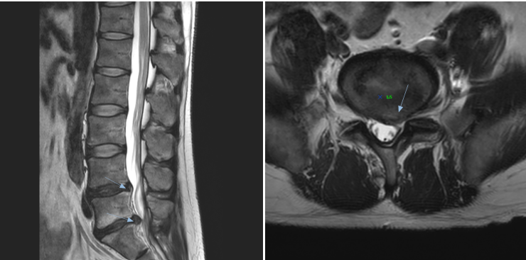
The next question is whether the nerve described on MRI is behaving like a symptomatic nerve during the clinical examination.
Why the MRI Wording Does Not Tell You Whether the Nerve Is Symptomatic
An MRI measures structure. An examination measures function. Those are two different questions, and only the second one is about how your nervous system is actually behaving.
Clinical findings that help establish whether a nerve root is involved include:
- sensory change in a distribution consistent with a specific root
- weakness in muscles supplied by that root
- a reduced or absent reflex on the affected side
- a radiating pain pattern that follows a plausible course
- nerve tension or provocation findings that reproduce the patient's familiar symptoms
Put simply: an MRI can show that something touches a nerve. The examination helps determine whether that nerve is actually behaving like an irritated or dysfunctional nerve.
What Is Radiculopathy?
Radiculopathy is a clinical diagnosis describing dysfunction of a spinal nerve root. It is established by the pattern of symptoms and by examination findings, most convincingly when sensory, motor and reflex findings agree on the same root.
Imaging supports the diagnosis when it shows something plausible at the matching level and side. It does not make the diagnosis by itself, and the word compression in a report is not a diagnosis of radiculopathy.
What Is Radicular Pain?
Radicular pain refers to pain generated by an irritated nerve root and felt along its course, often described as sharp, electric or lancinating down the leg. Radiculopathy specifically refers to demonstrable dysfunction of the root, such as numbness, weakness or reflex loss.
The two overlap and often occur together, but a person can have clear radicular pain with a normal neurological examination, and a person can have measurable root dysfunction with surprisingly little pain.
Why the Level and Side Matter
Correlation is the whole game. If a report describes a right-sided finding at L5-S1, the questions are whether your symptoms are on the right, whether they follow a distribution plausible for that root, and whether your examination findings point to the same place.
Dermatome charts are helpful for this, but they deserve a caution. Real dermatomal territories vary between people and overlap considerably, so a symptom pattern that does not match the textbook map exactly is not automatically wrong, and one that matches perfectly is not automatically proof. Clinicians weigh the whole picture, not one chart.
When Nerve Root Findings Need More Urgent Medical Evaluation
Most nerve root findings are handled without urgency. A few situations should prompt a same-day or urgent medical evaluation:
- weakness that is clearly progressing over days
- new loss of bowel or bladder control, or new difficulty urinating
- numbness in the saddle area, meaning the inner thighs, groin or buttocks
- severe neurological changes affecting both legs
- severe symptoms after significant trauma
- any finding your physician has flagged for prompt assessment
These are uncommon. Listing them is not a reason to read your report with dread, only a reason to know which symptoms should not wait.
For the surrounding terminology in your report, see the guide to reading a lumbar MRI report. If your report also describes narrowing of the nerve exit opening, that specific finding is covered in what neural foraminal narrowing means.
Frequently asked questions
References
- Fardon DF, Williams AL, Dohring EJ, et al. Lumbar disc nomenclature: version 2.0. Recommendations of the combined task forces of the North American Spine Society, the American Society of Spine Radiology and the American Society of Neuroradiology. Spine J. 2014;14(11):2525-2545. PMID: 24768732.
- Brinjikji W, Luetmer PH, Comstock B, et al. Systematic literature review of imaging features of spinal degeneration in asymptomatic populations. AJNR Am J Neuroradiol. 2015;36(4):811-816. PMID: 25430861.